|
CURRENT REFERENCES
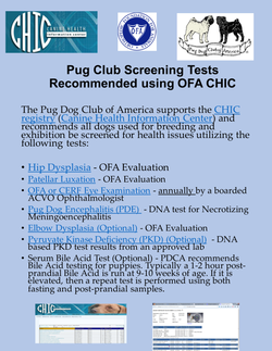
SCREENING TESTSThere is currently no method or test to screen Pugs for potential Pug Myelopathy neurological deficits. At this time, these tests for other inherited conditions are recommended by the Pug Dog Club of America.
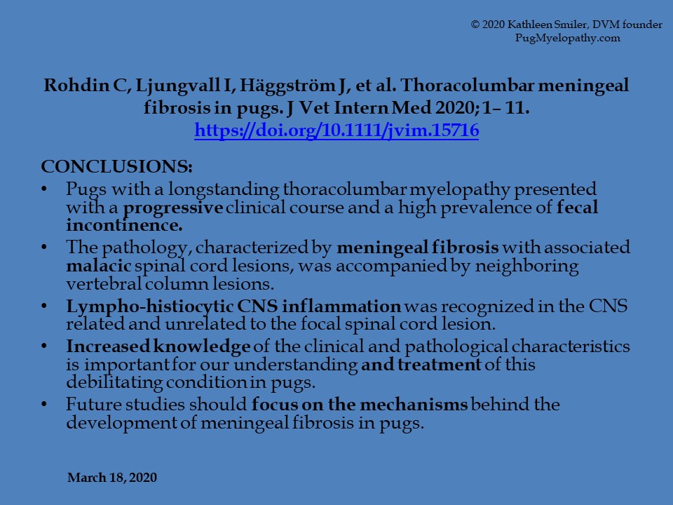
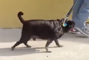
An Excellent Case History: My colleague, Michael Wong DVM, a board certified neurologist in Miami, FL has been extremely proactive in diagnosing and treating Pug spinal ataxia/paralysis. His "Case of the Month, Wellington," will help many individuals understand the characteristics and importance of this most significant cause of "weak rears" in middle aged Pugs. I cannot over emphasize the importance of seeking referral to a qualified veterinary neurologist or surgeon when symptoms first appear in affected animals. As Dr. Wong, ACVIM (Neurology) describes, there are multiple reasons Pugs may become ataxic, but in all cases, your dog needs to be evaluated promptly. Potential complications of urinary retention incontinence must be closely monitored. My sincere thanks to Dr. Wong for this excellent client education. ~Kathleen Smiler, DVM. |
TREATMENT OPTIONSMany veterinarians are not knowledgeable about this condition, as published information is not yet widely available. Pugs with rear limb ataxia may have had a previous diagnosis that was incomplete in light of this evolving information. Since so little is known, there is no consensus statement among neurologists about the best way to treat it. Surgery may be appropriate for individual cases, but it must be considered with interpretation of MRI imaging as soon as possible, after symptoms first occur, and may only delay progression of paralysis.... Click to Read More
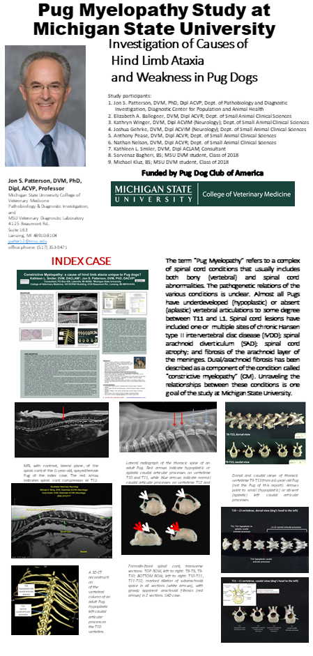
Pathogenesis of Pug Myelopathy "Hind limb ataxia or incoordination is common problem in Pugs—the second most important health issue in the breed, according to a survey of Pug owners and breeders by the Orthopedic Foundation for Animals (http://www.ofa.org/index.html ). In some cases, unsteadiness or incoordination in the hind limbs begins in middle-aged dogs (6-9 years old), but in most affected Pugs enrolled in the study at Michigan State University College of Veterinary Medicine (MSU CVM), the age of onset has been 9-12 years." Click to Read More by © Jon S. Patterson, DVM, PhD, Dipl, ACVP, Professor; Michigan State University College of Veterinary Medicine, Pathobiology & Diagnostic Investigation, and MSU Veterinary Diagnostic Laboratory, 4125 Beaumont Rd. Suite 163, Lansing, MI 48910-8104, patter12@msu.edu, Office phone: (517) 353-9471 Constrictive Myelopathy: a cause of hind limb ataxia unique to Pug dogs?
| ||||||
 Magnetic resonance imaging of the thoracolumbar spine of case no. 6, displaying extra-dural spinal cord compression associated with T11–12 disc protrusion and concurrent bilateral T11 caudal articular process aplasia. From left to right; T2-weighted mid sagittal of thoracolumbar spine, T2-weighted transverse at the affected level T11–12 (transverse level marked with a dashed line), three-dimensional reconstruction of thoracolumbar junction on CT (T11–12 marked with dashed line; the normal articular processes of T12 are highlighted with an arrow head). There is a marked focal increase in T2-weighted signal within the spinal cord despite only mild ventral extra-dural spinal cord compression (arrowed on transverse image)
- Research article Open Access Published: 31 May 2019
ABSTRACT
Background: A retrospective case series study was undertaken to describe the magnetic resonance imaging (MRI) findings in Pug dogs with thoracolumbar myelopathy and concurrent caudal articular process (CAP) dysplasia. Electronic clinical records were searched for Pug dogs who underwent MRI for the investigation of a T3-L3 spinal cord segment disease with subsequent confirmation of CAP dysplasia with computed tomography between January 2013 and June 2017. Clinical parameters age, gender, neuter status, body weight, urinary or faecal incontinence, severity and duration of clinical signs were recorded. MRI abnormalities were described. Univariable non-parametric tests investigated the association between the clinical parameters and evidence of extra- or intra-dural spinal cord compression on MRI.
Results: 18 Pug dogs were included. The median age was 106 months with median duration of clinical signs 5 months. All presented with variable severity of spastic paraparesis and ataxia; 50% suffered urinary/faecal incontinence. In all cases, MRI revealed a focal increase in T2-weighted signal intensity within the spinal cord at an intervertebral level where bilateral CAP dysplasia was present; this was bilateral aplasia in all but one case, which had one aplastic and one severely hypoplastic CAP. MRI lesions were associated with spinal cord compression in all but one case; intervertebral disc protrusion resulted in extra-dural compression in 10 (56%) cases; intra-dural compression was associated with a suspected arachnoid diverticulum in 4 (22%) cases and suspected pia-arachnoid fibrosis in 3 cases (17%). There was no association between clinical parameters and a diagnosis of intra-dural vs extra-dural compression. CAP dysplasia occurred at multiple levels in the T10–13 region with bilateral aplasia at T11–12 most often associated with corresponding spinal cord lesions on MRI.
Conclusions: All Pugs dogs in this study were presented for chronic progressive ambulatory paraparesis; incontinence was commonly reported. Although intervertebral disc disease was the most common radiologic diagnosis, intra-dural compression associated with arachnoid diverticulae/fibrosis was also common. Bilateral CAP aplasia was present in all but one Pug dog at the level of MRI detectable spinal cord lesions. A causal relationship between CAP dysplasia and causes of thoracolumbar myelopathy is speculated but is not confirmed by this study.

